
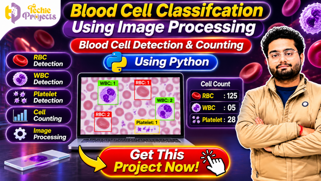
Looking for a practical Blood Cell Classification Using Image Processing project for your final-year engineering submission? Get a Python-based project package from Techie Projects and make your project development easier!
This project demonstrates how digital image processing techniques can identify and classify different blood cell types from microscopic images. It focuses on Red Blood Cells (RBCs), White Blood Cells (WBCs), and platelets using Python and computer vision concepts.
Instead of spending time developing everything from scratch, you can enquire about purchasing the project source code, documentation, implementation resources, and available technical support from Techie Projects.
Whether you are a B.Tech, BCA, MCA, or computer science engineering student, this project provides an opportunity to explore Python programming, image processing, computer vision, and biomedical image analysis.
📞 Contact Techie Projects for Project Enquiries
Want to purchase this project? Contact Techie Projects for pricing, source code availability, documentation, and implementation details.
- 📲 WhatsApp: https://wa.me/919460060699
- ☎️ Contact Number: +91 94600 60699
- 🌐 Website: https://techieprojects.com/
- 📸 Instagram: https://www.instagram.com/pythonprojects_/
🎬 Watch the Blood Cell Classification Using Image Processing Project Demo
Watch the project demonstration to understand how the Python application processes microscopic blood cell images, identifies different cell types, and displays detection and counting results.
🩸 What Is Blood Cell Classification Using Image Processing?
Blood Cell Classification Using Image Processing is a computer vision-based project that analyses microscopic blood smear images to identify different types of blood cells.
Human blood contains several important components, including:
- Red Blood Cells (RBCs): Responsible for carrying oxygen throughout the body.
- White Blood Cells (WBCs): Help the immune system fight infections.
- Platelets: Help the body form blood clots and control bleeding.
Manual examination of microscopic blood images can require time and specialised knowledge. Image processing provides a way to analyse digital images using computational techniques.
In this project, Python is used to load microscopic images, perform preprocessing, identify cell-like regions, and display classification results.
This project is designed for academic learning and demonstration. It is not a clinically validated medical diagnostic system.
🎯 Objectives of the Project
The main objective of Blood Cell Classification Using Image Processing is to demonstrate how Python can be used to analyse microscopic images and identify different blood cell categories.
The key objectives include:
- Load microscopic blood cell images into the application.
- Preprocess images to improve their quality.
- Identify relevant blood cell regions.
- Detect RBCs, WBCs, and platelets.
- Display detected cells using visual indicators.
- Calculate and display cell counts.
- Provide an understandable graphical interface.
⚙️ Key Features of Blood Cell Classification Using Image Processing
🔴 1. Red Blood Cell Detection
Red Blood Cells are among the most commonly observed components in microscopic blood images. The project demonstrates techniques for identifying RBC-like regions using image characteristics such as colour, shape, and boundaries.
🟣 2. White Blood Cell Detection
White Blood Cells have different visual characteristics from RBCs. Their appearance and staining patterns can help distinguish them in microscopic images.
The application demonstrates how image-processing operations can locate and highlight WBC-like regions.
🟡 3. Platelet Detection
Platelets are smaller blood components that can appear as small structures in microscopic images. The application demonstrates the identification and visualisation of platelet-like regions.
🔢 4. Blood Cell Counting
Counting is an important part of image analysis. After detecting cell regions, the application can display the number of identified objects in the processed image.
🖼️ 5. Image Processing and Visualisation
The project displays processed images and highlights detected regions using visual indicators. This helps users understand the difference between original and processed images.
💻 6. User-Friendly Interface
A graphical interface allows users to select images, run the analysis, and view the output.
🌟 Benefits of Blood Cell Classification Using Image Processing
Blood Cell Classification Using Image Processing offers several benefits for students exploring Python programming and computer vision. It combines theoretical concepts with practical implementation and provides an opportunity to understand how digital images can be analysed.
Some important benefits include:
- Automated Image Analysis: Demonstrates how computers can analyse microscopic images.
- Cell Detection: Shows techniques for identifying different blood cell categories.
- Python Programming: Provides practical experience with Python libraries.
- Visual Results: Displays detected cell regions and counting information.
- Academic Learning: Helps students understand image preprocessing, segmentation, and feature extraction.
- Future Enhancements: Provides a foundation for exploring deep learning and advanced classification techniques.
🔬 Why Is Blood Cell Classification Using Image Processing Important?
Blood Cell Classification Using Image Processing demonstrates how computer vision techniques can be applied to biological image analysis. Microscopic images contain different visual patterns, and image-processing algorithms can help identify and analyse these patterns.
By working on this project, students can understand how image enhancement, segmentation, contour detection, and feature extraction contribute to an image-analysis workflow.
The project also provides practical exposure to OpenCV and NumPy, which are widely used in Python-based computer vision applications.
💻 How to Develop Blood Cell Classification Using Image Processing?
Developing Blood Cell Classification Using Image Processing involves combining several Python programming and image-analysis techniques.
First, collect suitable microscopic blood cell images for experimentation. Next, load the images using OpenCV and apply preprocessing operations to improve their quality. After preprocessing, use segmentation techniques to separate relevant regions from the background.
The next stage involves extracting useful image features and applying the implemented classification logic. Finally, display the detected regions and counting results through the application interface.
Students can further improve the project by experimenting with different datasets, segmentation approaches, and deep learning models.
🧠 Technologies Used in the Project
The project uses Python and image-processing concepts to analyse microscopic blood cell images.
| Technology | Purpose |
|---|---|
| Python | Main programming language |
| OpenCV | Image processing and computer vision |
| NumPy | Numerical operations and image arrays |
| Tkinter | Graphical interface, if included |
| Matplotlib | Image visualisation, if included |
🐍 Python
Python is widely used in machine learning, data science, automation, and computer vision. Its extensive library ecosystem makes it suitable for developing image-processing applications.
👁️ OpenCV
OpenCV is a popular computer vision library. It provides functions for image reading, resizing, colour conversion, filtering, thresholding, and contour detection.
🔢 NumPy
NumPy provides support for multidimensional arrays and numerical calculations. Digital images can be represented as arrays of pixel values, making NumPy useful for image manipulation.
🔄 How Does the Project Work?
The Blood Cell Classification Using Image Processing application follows a sequence of image-processing operations.
Step 1: 📂 Upload a Blood Cell Image
The user selects a microscopic blood smear image from the computer. The application loads the image for further processing.
Step 2: 🖼️ Image Preprocessing
The input image may contain noise, uneven lighting, or unwanted visual details. Preprocessing techniques can improve the image before detection.
These techniques may include:
- Image resizing
- Noise reduction
- Colour space conversion
- Contrast enhancement
- Image filtering
Step 3: 🎨 Image Segmentation
Segmentation separates relevant image regions from the background. Thresholding and colour-based processing can help isolate cell-like structures.
Step 4: 🔍 Feature Extraction
After segmentation, the application can examine visual properties of identified regions.
Common features include:
- Area
- Perimeter
- Shape
- Colour
- Contour
- Region size
Step 5: 🧬 Cell Classification
The identified regions are analysed using the project’s implemented classification logic. The application aims to distinguish RBC-like, WBC-like, and platelet-like regions.
Step 6: 🔢 Cell Counting
The application counts detected regions and presents the results. The output may include separate counts for different cell categories, depending on the implementation.
Step 7: 📊 Display Final Results
Finally, the application displays the processed image and classification results for review.
🏥 Applications of Blood Cell Image Processing
Image processing has several potential applications in biological image analysis and academic research.
🔬 1. Microscopic Image Analysis
Computer vision techniques can be used to study structures visible in microscopic images.
🧪 2. Biomedical Engineering Education
Students can explore how programming and image-processing techniques are applied to healthcare-related problems.
🎓 3. Academic Research
The project can serve as a starting point for experiments involving segmentation, feature extraction, and object classification.
🤖 4. Computer Vision Learning
Students can learn how digital images are represented and how algorithms identify visual patterns.
📚 5. Image Processing Demonstrations
The application provides a practical example of applying image-processing concepts to a real-world-inspired dataset.
🌟 Why Choose This Project for Your Final Year?
Choosing the right final-year project is important for demonstrating technical skills and completing academic requirements.
Blood Cell Classification Using Image Processing offers several learning opportunities.
- Practical Python Implementation: Understand how Python libraries are used to develop image-processing applications.
- Real-World Inspired Problem: Connect computer vision concepts with microscopic image analysis.
- Multiple Technical Concepts: Explore preprocessing, segmentation, feature extraction, classification, and counting.
- Demonstrable Output: Review visual results and explain the implementation during your project presentation.
- Future Development: Explore deep learning, CNN-based classification, and improved image segmentation.
📦 What Do You Get When You Purchase This Project from Techie Projects?
If you are interested in purchasing this project, contact Techie Projects to confirm the available package and deliverables.
Depending on the selected package, the project may include:
- Python project source code
- Required project files
- Dataset or sample images, where available
- Installation and execution guidance
- Project documentation, where included
- Demonstration resources
- Technical support options, as agreed
Before placing an order, confirm the exact deliverables, software requirements, support duration, and additional services.
💻 Software and Hardware Requirements
The following are general requirements for running a Python-based image-processing application. Actual requirements depend on the delivered implementation.
Software Requirements
- Operating System: Windows 10 or Windows 11
- Programming Language: Python
- Image Processing Library: OpenCV
- Numerical Library: NumPy
- Development Environment: VS Code, PyCharm, or another Python IDE
Hardware Requirements
- Processor: Intel or AMD processor
- RAM: 4 GB minimum recommended; 8 GB or more preferable
- Storage: Sufficient free disk space
- Input Devices: Keyboard and mouse
- Display: Standard computer monitor
🔮 Future Scope of the Project
The project can be extended using more advanced computer vision and artificial intelligence techniques.
🤖 1. Deep Learning-Based Classification
CNN models can be explored to classify blood cell images using labelled datasets.
📊 2. Improved Detection Accuracy
Better image preprocessing, segmentation, and feature extraction methods can be evaluated to improve results.
☁️ 3. Web-Based Application
The project could be extended into a web application where users upload images and view processing results through a browser.
📱 4. Mobile Application
A mobile interface could provide a convenient way to access image-processing functionality.
🧠 5. Advanced Image Analysis
Further research could explore cell morphology, image quality assessment, and more detailed classification methods.
🎓 Who Can Purchase This Project?
This project may be useful for students and learners from several academic backgrounds:
- B.Tech Computer Science Engineering students
- B.Tech Information Technology students
- BCA students
- MCA students
- M.Tech students
- Diploma engineering students
- Students learning Python and OpenCV
- Learners interested in computer vision and biomedical image processing
Students should understand, test, and adapt the implementation according to their institution’s academic requirements.
❓ Frequently Asked Questions (FAQs)
1. What is Blood Cell Classification Using Image Processing?
It is a Python-based computer vision project that analyses microscopic blood images and demonstrates the identification and counting of RBCs, WBCs, and platelets.
2. Which programming language is used?
Python is used as the main programming language.
3. Which library is used for image processing?
OpenCV is a commonly used library for image processing and computer vision in this type of project.
4. Can I use this project for my final-year submission?
It can be considered as an academic project topic. Check your institution’s requirements and ensure that you understand and can explain the implementation.
5. Does the project detect RBC, WBC, and platelets?
The project is designed to demonstrate detection and classification of these cell categories. Actual performance depends on the implementation and input images.
6. Does the project include source code?
Contact Techie Projects to confirm the available package and source-code deliverables.
7. Can the project be extended using deep learning?
Yes. CNN-based classification and other deep learning techniques can be explored as future enhancements.
8. How can I purchase this project?
Contact Techie Projects through WhatsApp at +91 94600 60699 or visit https://techieprojects.com/.
9. Is this project a medical diagnostic tool?
No. It is an educational and demonstration project. It is not clinically validated and should not be used for medical diagnosis or treatment decisions.
🔗 Explore Related Python and Image Processing Projects
If you are interested in similar academic projects, explore other project categories available at Techie Projects:
- 🧠 Artificial Intelligence Projects
- 👁️ Image Processing Projects
- 🤖 Machine Learning Projects
- 📊 Data Science Projects
- 🐍 Python Projects
- 🧬 Deep Learning Projects
- 💻 Computer Vision Projects
Visit www.techieprojects.com to explore more project ideas and implementation resources.
🚀 Purchase Blood Cell Classification Using Image Processing Project from Techie Projects
Want to work on Blood Cell Classification Using Image Processing without developing the entire application from the beginning?
Techie Projects provides academic project resources for students interested in Python, machine learning, artificial intelligence, and image processing.
Contact us to enquire about the available project package, source code, documentation, installation guidance, and technical support.
📲 WhatsApp: https://wa.me/919460060699
🌐 Website: https://techieprojects.com/
☎️ Contact: +91 94600 60699