
Introduction
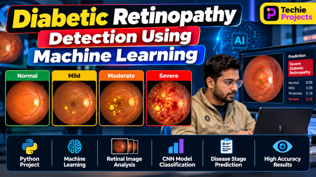
Diabetic retinopathy detection using machine learning is an important computer vision project that demonstrates how artificial intelligence can analyze retinal images to identify signs associated with diabetic eye disease. Diabetic retinopathy is a complication of diabetes that can damage the blood vessels in the retina and may lead to vision loss if not diagnosed and managed appropriately.
Traditional retinal image examination requires trained medical professionals. Machine learning and deep learning techniques can help researchers develop systems that analyze retinal fundus images and classify them according to patterns associated with different stages of diabetic retinopathy.
This Diabetic Retinopathy Detection Using Machine Learning project uses Python, image processing, and deep learning concepts to demonstrate how an automated retinal image classification system can be developed. It is suitable for B.Tech students, computer science engineering students, and learners interested in artificial intelligence, machine learning, and medical image analysis.
Important: This project is intended for educational and research purposes. It is not a substitute for professional eye examinations or a clinically validated diagnostic system.
🎬 Project Working Demo
Watch the project demonstration to understand how the Diabetic Retinopathy Detection Using Machine Learning system works in practice.
The demo can showcase how a retinal fundus image is uploaded, preprocessed, and passed to a trained machine learning model for classification. It can also demonstrate how the predicted category and model output are displayed in the application.
What Is Diabetic Retinopathy Detection?
Diabetic retinopathy detection is the process of analyzing retinal images to identify visual patterns that may indicate diabetic retinopathy. These patterns can include microaneurysms, hemorrhages, and other retinal abnormalities.
A machine learning model learns patterns from labeled retinal images and uses those patterns to predict a class for a new image.
Depending on the dataset and implementation, the system may classify images into five categories:
- No DR: No diabetic retinopathy detected in the labeled image.
- Mild: Features associated with mild non-proliferative diabetic retinopathy.
- Moderate: Features associated with moderate diabetic retinopathy.
- Severe: Features associated with severe non-proliferative diabetic retinopathy.
- Proliferative DR: Features associated with proliferative diabetic retinopathy.
The number and names of classes depend on the dataset being used. The model’s predictions should not be treated as a confirmed medical diagnosis.
Objectives of the Project
The main objectives of this Python project are:
- To understand how machine learning can be applied to retinal image analysis.
- To preprocess retinal fundus images for model training.
- To develop an image classification model using a suitable machine learning or deep learning approach.
- To classify retinal images into the categories supported by the selected dataset.
- To evaluate model performance using appropriate metrics.
- To build a simple interface for demonstrating predictions.
- To explore the practical applications and limitations of AI-assisted medical image analysis.
Key Features of the Diabetic Retinopathy Detection System
The following features can be included, depending on the project implementation.
1. Retinal Image Upload
Users can select a retinal fundus image from a local computer. The application loads the image and prepares it for processing.
2. Image Preprocessing
Images are resized to the input dimensions expected by the model. Additional preprocessing may include pixel normalization, image enhancement, and data augmentation during training.
3. Deep Learning-Based Classification
A convolutional neural network (CNN) can learn useful visual features from retinal images. Alternatively, a pretrained model can be fine-tuned using transfer learning.
4. Prediction Display
The application displays the predicted class and, if implemented, the model’s class probability scores. These scores represent the model’s output, not a guaranteed measure of clinical certainty.
5. Performance Evaluation
The trained model can be evaluated using a separate test dataset to measure its performance on images that were not used for training.
6. User-Friendly Interface
A simple interface developed with Streamlit, Flask, or Tkinter can make the project easier to demonstrate.
Technologies Used
The technologies depend on the chosen implementation. A typical project may use:
- Programming Language: Python
- Image Processing: OpenCV or Pillow
- Numerical Operations: NumPy
- Data Analysis: Pandas
- Deep Learning: TensorFlow/Keras or PyTorch
- Visualization: Matplotlib or Seaborn
- Application Interface: Streamlit, Flask, or Tkinter
- Development Environment: Jupyter Notebook, Google Colab, or VS Code
Students should select compatible library versions and document the environment used to train and run the model.
Dataset for Diabetic Retinopathy Detection
A labeled retinal image dataset is required to train and evaluate the model. Public research datasets, such as the APTOS 2019 Blindness Detection dataset and EyePACS-based datasets, are commonly used for educational experiments in diabetic retinopathy classification.
Before selecting a dataset, review its license, permitted uses, class labels, image quality, and data distribution.
A typical dataset workflow includes:
- Collect or download an appropriately licensed dataset.
- Inspect the image files and corresponding labels.
- Check the number of images in each class.
- Split the dataset into training, validation, and test sets.
- Apply preprocessing consistently.
- Train the model using the training data.
- Tune the model using validation data.
- Evaluate final performance on the held-out test data.
Avoid placing near-duplicate images or images from the same patient across different splits where patient-level identifiers are available. Data leakage can make evaluation results look better than real-world performance.
Working of the Diabetic Retinopathy Detection Project
The system follows a series of steps to transform retinal images into model predictions.
Step 1: Load the Dataset
Load the retinal images and their corresponding labels. Verify that image paths and labels match correctly.
Step 2: Preprocess the Images
Resize the images to a consistent input size and apply suitable normalization. Image enhancement or augmentation may be used when appropriate for the selected dataset.
Step 3: Split the Data
Create training, validation, and test sets. Keep the test set separate until final evaluation to obtain a more reliable estimate of model performance.
Step 4: Build the CNN Model
A CNN can be designed using convolutional layers, activation functions, pooling layers, and fully connected layers. For a transfer-learning approach, a pretrained image classification model can be adapted to the dataset’s classes.
Step 5: Train the Model
Train the model on labeled retinal images. Monitor training and validation metrics to identify overfitting and determine whether adjustments to the model or training process are needed.
Step 6: Evaluate the Model
Evaluate the final model on the test set. Useful metrics include accuracy, precision, recall, F1-score, confusion matrix, and per-class performance.
For medical image classification, overall accuracy alone may not be sufficient. Recall and sensitivity for relevant classes, false-negative rates, and performance across different patient groups are also important considerations.
Step 7: Predict on a New Image
Load a new retinal image, apply the same preprocessing used during training, and pass it to the trained model. Display the predicted category and any relevant output scores.
Step 8: Display the Results
Present the result in a clear interface with an educational disclaimer. The application should explain that its output is experimental and requires professional clinical assessment.
CNN Architecture for Retinal Image Classification
A convolutional neural network is commonly used for image classification because it can learn spatial patterns from images.
A basic CNN architecture may contain:
- Convolutional layers: Extract visual features such as edges, textures, and patterns.
- Activation functions: Introduce non-linearity into the network.
- Pooling layers: Reduce the spatial dimensions of feature maps.
- Dropout layers: Help reduce overfitting during training.
- Fully connected layers: Combine learned features for classification.
- Output layer: Produces scores for the supported classes.
The architecture should be selected according to dataset size, computing resources, and evaluation results. A more complex model does not automatically produce better or more reliable predictions.
Software Requirements
A typical software setup includes:
- Python 3
- TensorFlow/Keras or PyTorch
- NumPy and Pandas
- OpenCV or Pillow
- Matplotlib
- Jupyter Notebook or VS Code
- Streamlit, Flask, or Tkinter if a graphical interface is required
Install compatible versions of the libraries in a virtual environment to reduce dependency conflicts.
Hardware Requirements
The project can generally be developed on a standard computer with:
- A dual-core or better processor
- At least 8 GB RAM recommended for comfortable experimentation
- Sufficient storage for the dataset, model files, and results
- An optional NVIDIA GPU for faster training
A GPU is not mandatory for every implementation. Smaller experiments can run on a CPU, while larger datasets and deep learning models may require more memory and training time.
Advantages of the Project
- Demonstrates a practical application of machine learning and computer vision.
- Helps students understand image preprocessing and classification.
- Introduces CNNs and transfer learning.
- Provides experience with dataset preparation and model evaluation.
- Can be extended with a web interface and visual reports.
- Offers an opportunity to study the role and limitations of AI in medical image analysis.
Limitations and Challenges
Dataset Quality
Blurry, poorly illuminated, or inconsistently labeled images can affect model performance.
Class Imbalance
Some severity categories may contain fewer examples than others. Class imbalance can cause the model to perform unevenly across categories.
Generalization
A model trained on one dataset may perform differently on images collected using other cameras, clinical settings, or patient populations.
False Predictions
The system may incorrectly classify an image, including missing signs of disease. Therefore, it should not be used as a standalone clinical diagnostic tool.
Computing Requirements
Training time and memory usage depend on dataset size, model architecture, image resolution, and available hardware.
Applications of Diabetic Retinopathy Detection
Research and educational applications include:
- Medical image classification experiments
- Computer vision and deep learning demonstrations
- AI research on retinal image analysis
- Student projects exploring automated image assessment
- Prototype interfaces for demonstrating model predictions
- Studies of model accuracy, fairness, and generalization
Any clinical application requires appropriate validation, qualified medical oversight, and compliance with applicable regulations.
Future Scope
The project can be expanded in several ways:
- Experiment with transfer learning using pretrained CNN architectures.
- Add explainability methods, such as Grad-CAM, to visualize image regions influencing predictions.
- Improve class imbalance handling and compare training strategies.
- Develop a Streamlit or Flask interface for uploading images and viewing results.
- Compare multiple architectures using the same data splits and evaluation metrics.
- Test generalization on an independent dataset.
- Add confidence calibration and clearer uncertainty reporting.
- Conduct fairness and robustness evaluations across different image sources and patient groups.
These improvements can make the project more useful for research and learning, but they do not by themselves establish clinical safety.
Frequently Asked Questions (FAQs)
1. What is diabetic retinopathy detection using machine learning?
It is an image classification task in which a trained model analyzes retinal images to predict categories associated with diabetic retinopathy.
2. Which programming language is used for this project?
Python is commonly used because it provides libraries for image processing, data analysis, deep learning, and application development.
3. Which algorithm is suitable for diabetic retinopathy detection?
CNNs and pretrained deep learning models using transfer learning are common choices. The best approach depends on the dataset, available resources, and measured performance.
4. Is a GPU required to develop this project?
No. Smaller models can be trained on a CPU, although a compatible GPU can significantly reduce training time for larger experiments.
5. Can this project be used as a final-year engineering project?
Yes. It can be an educational final-year project for students interested in Python, machine learning, deep learning, and computer vision. Students should understand the implementation, dataset, evaluation, and limitations before presenting their work.
6. Can the model diagnose diabetic retinopathy accurately in real patients?
A project’s performance on a public dataset does not establish clinical accuracy. Clinical use requires independent validation, appropriate medical oversight, and compliance with applicable requirements.
7. Which evaluation metrics should be used?
Accuracy, precision, recall, F1-score, a confusion matrix, and class-specific sensitivity can help assess model performance. The choice of metrics should reflect the intended research question and the consequences of different prediction errors.
🎓 Explore Related Machine Learning Projects
If you are interested in similar Python and AI projects, explore these related topics on Techie Projects:
- Machine Learning Projects Using Python
- Deep Learning Projects for Final-Year Students
- Image Processing Projects Using Python
- Skin Cancer Detection Using Machine Learning
- Dental Caries Detection Using Deep Learning
- Sign Language Recognition Using Machine Learning
Link each topic to its relevant page on the Techie Projects website.
🚀 Need Help With Your Final-Year Project?
Looking for a Python, machine learning, deep learning, or computer vision project?
Techie Projects can help you explore project ideas, implementation options, software requirements, and documentation support.
- 📲 Call/WhatsApp: +91-9460060699
- 🌎 Website: www.techieprojects.com
- 📺 Instagram: @pythonprojects_
Contact us to discuss your academic requirements and available project support.
Conclusion
Diabetic retinopathy detection using machine learning is a valuable educational project for understanding how deep learning can be applied to retinal image classification. By preparing a labeled dataset, preprocessing images, training a CNN or transfer-learning model, and evaluating its predictions, students can explore important concepts in Python, machine learning, and medical computer vision.
The project can be extended through model comparisons, explainability techniques, and a user-friendly interface. However, experimental model predictions should not be treated as a medical diagnosis. Reliable clinical use requires much more extensive validation and professional oversight.