
Introduction
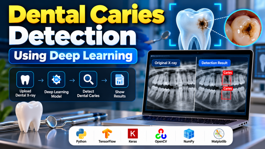
Dental Caries Detection Using Deep Learning is an artificial intelligence and computer vision project designed to identify possible dental caries, commonly known as tooth decay or cavities, from dental images such as X-rays. The project uses deep learning techniques to analyze dental images and learn visual patterns associated with caries.
Traditional dental examination relies on clinical examination and dental imaging performed by qualified dental professionals. A deep learning system can be developed as an educational decision-support prototype that analyzes images and highlights regions that may require further examination.
This project is suitable for B.Tech, BCA, MCA, Artificial Intelligence, Machine Learning, and Computer Science students interested in medical image analysis and deep learning.
Important: This project is for educational and research purposes only. A machine learning prediction should not be considered a medical diagnosis or a replacement for examination by a qualified dentist.
What Is Dental Caries?
Dental caries is a condition in which the hard tissues of a tooth are gradually damaged. It is commonly referred to as tooth decay or cavities.
Dental caries may appear as characteristic changes in dental images. Detecting these changes manually can require experience and careful examination.
Deep learning can be explored as a computer-aided approach for analyzing dental images and identifying patterns associated with possible caries.
What Is Dental Caries Detection Using Deep Learning?
Dental caries detection using deep learning involves training an image-analysis model using labeled dental images.
The model learns visual patterns from the training dataset and can subsequently analyze a new image to produce a prediction.
A simplified workflow is:
Dental X-ray → Image Preprocessing → Deep Learning Model → Feature Extraction → Caries Classification/Detection → Result
Depending on the dataset and model architecture, the system can perform:
- Image classification
- Caries/non-caries classification
- Multi-class classification
- Object detection
- Region localization
- Segmentation
Objectives of the Project
The major objectives of the Dental Caries Detection Using Deep Learning project are:
- To understand medical image analysis using deep learning.
- To preprocess dental X-ray images for model training.
- To develop an image classification or detection model.
- To identify image patterns associated with possible dental caries.
- To evaluate the performance of the trained model.
- To demonstrate the use of artificial intelligence in dental image analysis.
- To develop an educational prototype for computer-aided dental image analysis.
How Dental Caries Detection Works
The project can be divided into several stages.
1. Dataset Collection
The first step is to collect an appropriate dataset containing dental images.
The dataset should contain correctly labeled images, for example:
- Caries
- No caries
For more advanced projects, the dataset may contain annotations showing the location of suspected carious lesions.
The quality and diversity of the dataset have a significant effect on model performance.
2. Image Preprocessing
Dental images may have different resolutions, sizes, and brightness levels. Therefore, preprocessing is performed before feeding them into the deep learning model.
Common preprocessing operations include:
- Image resizing
- Pixel normalization
- Noise reduction
- Contrast adjustment
- Image format conversion
- Data augmentation
Data augmentation can create variations of training images using transformations such as:
- Rotation
- Horizontal flipping where clinically appropriate
- Zooming
- Cropping
- Translation
- Brightness adjustments
The selected transformations should preserve clinically relevant image characteristics.
3. Deep Learning Model
A convolutional neural network (CNN) can be used to learn visual features from dental images.
A CNN automatically learns hierarchical features from images.
For example:
Input Image → Convolution Layers → Feature Maps → Pooling → Dense Layers → Prediction
For transfer-learning-based implementations, a pre-trained architecture can also be used.
Possible architectures include:
- ResNet
- EfficientNet
- MobileNet
- DenseNet
- VGG
- Inception
The most suitable architecture depends on the dataset, task, computational resources, and evaluation results.
4. Model Training
The preprocessed images are divided into suitable subsets, such as:
- Training set
- Validation set
- Test set
The model learns from the training images while the validation set can be used to monitor performance during training.
Important training parameters include:
- Learning rate
- Batch size
- Number of epochs
- Optimizer
- Loss function
For binary classification, a suitable binary classification loss function can be used. For multiple classes, an appropriate multi-class loss function can be selected.
5. Model Evaluation
After training, the model should be evaluated using an independent test dataset.
Useful evaluation metrics include:
Accuracy
Measures the proportion of correct predictions.
Precision
Measures how many predicted positive cases are actually positive.
Recall
Measures how many actual positive cases are correctly identified.
F1-Score
Combines precision and recall into a single metric.
Confusion Matrix
A confusion matrix helps visualize correct and incorrect predictions across classes.
For medical-image applications, accuracy alone should not be treated as sufficient. Metrics such as sensitivity/recall, specificity, precision, and appropriate validation are important.
Dental Caries Detection Project Workflow
The complete workflow can be represented as:
Dental Image Dataset
↓
Image Preprocessing
↓
Data Augmentation
↓
Train/Test Data Preparation
↓
CNN or Transfer Learning Model
↓
Model Training
↓
Model Evaluation
↓
Save Trained Model
↓
Upload New Dental Image
↓
Model Prediction
↓
Display Result
Key Features
A dental caries detection project can include the following features:
- Dental image upload
- Image preprocessing
- Deep learning-based analysis
- Caries classification
- Prediction probability/confidence
- Detection-result visualization
- Model performance evaluation
- Simple web interface
For an advanced version, the application can display the approximate region detected by an object detection or segmentation model.
Technologies Used
| Technology | Purpose |
|---|---|
| Python | Project development |
| TensorFlow/Keras | Deep learning |
| OpenCV | Image processing |
| NumPy | Numerical operations |
| Pandas | Data handling |
| Matplotlib | Visualization |
| CNN | Image feature extraction |
| Transfer Learning | Reusing pre-trained visual features |
A different deep learning framework or model architecture can also be used depending on the project implementation.
Software Requirements
The following software can be used:
- Windows, Linux, or macOS
- Python 3.x
- TensorFlow/Keras
- OpenCV
- NumPy
- Pandas
- Matplotlib
- Jupyter Notebook or VS Code
For deployment, the project can also use frameworks such as Streamlit, Flask, or FastAPI.
Hardware Requirements
Recommended hardware includes:
- Intel or AMD processor
- Minimum 8 GB RAM
- 10 GB or more available storage
- NVIDIA GPU recommended for faster deep learning training
- Stable internet connection for downloading datasets and libraries
A GPU is not mandatory for small experiments, but it can significantly reduce training time.
Applications
Deep learning-based dental image analysis can have potential applications in:
- Dental image research
- Computer-aided image analysis
- Medical AI research
- Dental education
- Academic deep learning projects
- Medical image classification research
- Dental screening research prototypes
Any real-world clinical application requires appropriate validation, regulatory consideration, professional oversight, and evaluation on representative clinical data.
Advantages of the Project
The project provides several educational advantages:
- Demonstrates practical deep learning
- Introduces medical image analysis
- Provides experience with CNN architectures
- Can use transfer learning
- Supports image classification experiments
- Can be extended to object detection
- Provides experience with model evaluation
- Can be deployed as a web application
Limitations
Dental caries detection using deep learning also has important limitations.
Dataset Quality
Poor-quality or incorrectly labeled images can reduce model performance.
Limited Dataset
A model trained on a small dataset may not generalize well to images from different sources.
Image Quality
Blurred, noisy, or poorly exposed images can affect predictions.
Similar Visual Patterns
Other dental conditions or image artifacts may have visual characteristics that can confuse a model.
Clinical Validation
A model developed as a student project should not be assumed to be clinically reliable without appropriate validation.
Future Scope
The project can be enhanced in several ways.
Object Detection
Instead of only classifying an image, an object detection model can be trained to highlight regions associated with suspected caries.
Image Segmentation
Segmentation can be explored to identify specific regions of interest at the pixel level.
Transfer Learning
Pre-trained models such as ResNet, EfficientNet, DenseNet, or MobileNet can be evaluated to compare performance.
Explainable AI
Techniques such as Grad-CAM can be explored to visualize areas that influenced the model’s prediction.
Web Application
The trained model can be integrated into a Streamlit, Flask, or FastAPI application where users can upload an image and view the model output.
Larger Dataset
Using a larger and more diverse dataset can help evaluate whether the model generalizes across different image sources and patient populations.
Frequently Asked Questions
What is dental caries detection using deep learning?
Dental caries detection using deep learning is an image-analysis approach in which a trained neural network analyzes dental images to classify or identify patterns associated with possible tooth decay.
Can Python be used for dental caries detection?
Yes. Python libraries such as TensorFlow, Keras, OpenCV, NumPy, and Matplotlib can be used to develop an educational dental image-analysis project.
Which deep learning model can be used?
CNN architectures such as ResNet, EfficientNet, DenseNet, MobileNet, VGG, and Inception can be evaluated depending on the dataset and project requirements.
Is this suitable as a final-year project?
Yes. Dental caries detection is a suitable academic project for students interested in artificial intelligence, deep learning, computer vision, and medical image analysis.
Can the project detect the exact location of dental caries?
A basic classification model generally predicts a class for the entire image. To identify specific regions, an object detection or segmentation model with suitable annotations is required.
Can this model be used for medical diagnosis?
A student or research prototype should not be used as a standalone medical diagnostic tool. Clinical diagnosis should be performed by qualified dental professionals using appropriate clinical examination and diagnostic procedures.
Conclusion
Dental Caries Detection Using Deep Learning is an interesting computer vision project that demonstrates how artificial intelligence can be applied to dental image analysis. By using CNNs or transfer learning, a model can be trained to recognize patterns associated with possible dental caries in appropriately labeled images.
The project gives students practical experience with Python, deep learning, image preprocessing, CNNs, transfer learning, model evaluation, and medical image analysis. It can also be extended into advanced areas such as object detection, segmentation, explainable AI, and web-based deployment.